A Matter of Life and Cell Death
MOLECULAR VISUALIZATION, EDITORIAL
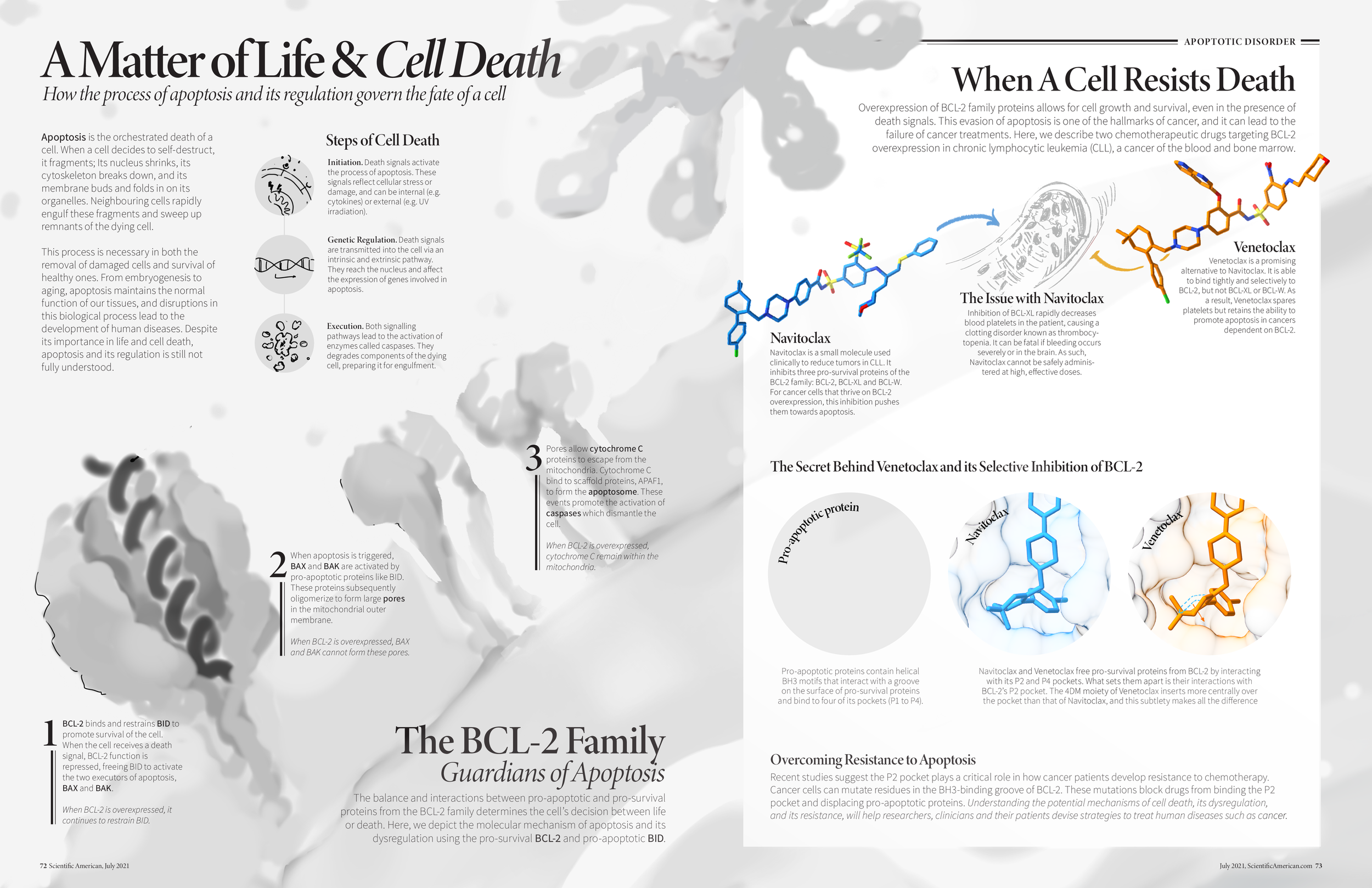
Apoptosis is a cellular process vital for human function. This spread illustrates how the overexpression of a particular protein alters apoptosis and leads to the development of cancer. It also visualizes the mechanism of a drug used to improve clinical outcomes.
Adobe Illustrator; Adobe Photoshop; Autodesk Maya; Protein Imager; UCSF Chimera; VMD
TOOLS
RESEARCH AND IDEATION
Fascinated by death, I centered my story on apoptosis and its dysregulation. I first conducted a media audit on a protein family of interest, the pro-survival BCL-2. This gave me an idea of what worked well in existing communication products and what could be improved upon.
I created thumbnails to plan the flow and visual style of the piece. I went with a story that provided an overview of apoptosis and its role in human cancer.
PRE-PRODUCTION
I had unanswered questions regarding this story, so I researched and compiled a list of resources to fill my gaps in knowledge. I started writing copy with the help of this list — this helped me create a comprehensive draft. As I refined the layout, I cut down on the depth of the story until I was satisfied with the text-visual balance.
STYLE TEST
I experimented with the visual treatment of molecules using Protein Imager and UCSF Chimera. I decided to create a cellular landscape with Van der Waals surface rendering to illustrate the environment in which apoptosis occurs. A different treatment, toon-shading, was used to illustrate the drug mechanism and separate the two halves of the story.
PRODUCTION
Molecular data from multiple sources (PDB, journal articles, UniProt, and PubChem) were integrated to create the 3D cellscape in Maya and 2D assets in Protein Imager. These were then colorized in Photoshop and compiled in Illustrator.
Birkinshaw, R.W., Gong, Jn., Luo, C.S. et al. (2019). Structures of BCL-2 in complex with venetoclax reveal the molecular basis of resistance mutations. Nat Commun, 10, 2385. https://doi.org/10.1038/s41467-019-10363-1
Birkinshaw, R. W., Iyer, S., Lio, D., Luo, C. S., Brouwer, J. M., Miller, M. S., Robin, A. Y., Uren, R. T., Dewson, G., Kluck, R. M., Colman, P. M., & Czabotar, P. E. (2021). Structure of detergent-activated BAK dimers derived from the inert monomer. Molecular Cell, 81(10). https://doi.org/10.1016/j.molcel.2021.03.014
Hanahan, D., & Weinberg, R. A. (2011). Hallmarks of Cancer: The Next Generation. Cell, 144(5), 646–674. https://doi.org/10.1016/j.cell.2011.02.013
Horvath, S. E., & Daum, G. (2013). Lipids of mitochondria. Progress in Lipid Research, 52(4), 590-614. doi:10.1016/j.plipres.2013.07.002
Kønig, S. M., Rissler, V., Terkelsen, T., Lambrughi, M., & Papaleo, E. (2019). Alterations of the interactome of Bcl-2 proteins in breast cancer at the transcriptional, mutational and structural level. PLOS Computational Biology, 15(12). https://doi.org/10.1371/journal.pcbi.1007485
Kühlbrandt W. (2015). Structure and function of mitochondrial membrane protein complexes. BMC biology, 13, 89. https://doi.org/10.1186/s12915-015-0201-x
Mannella, C. A. (2004). Mitochondrial Membranes, Structural Organization. Encyclopedia of Biological Chemistry, 720–724. https://doi.org/10.1016/b0-12-443710-9/00046-6
Pfanner N, Warscheid B, Wiedemann N. Mitochondrial proteins: from biogenesis to functional networks. Nat Rev Mol Cell Biol. 2019 May;20(5):267-284. doi: 10.1038/s41580-018-0092-0. Erratum in: Nat Rev Mol Cell Biol. 2021 May;22(5):367. PMID: 30626975; PMCID: PMC6684368.
Renehan, A. G., Booth, C., & Potten, C. S. (2001). What is apoptosis, and why is it important?. BMJ (Clinical research ed.), 322(7301), 1536–1538. https://doi.org/10.1136/bmj.322.7301.1536
Singh, R., Letai, A. & Sarosiek, K. (2019). Regulation of apoptosis in health and disease: the balancing act of BCL-2 family proteins. Nat Rev Mol Cell Biol, 20, 175–193. https://doi.org/10.1038/s41580-018-0089-8
Tahir, S. K., Smith, M. L., Hessler, P., Rapp, L. R., Idler, K. B., Park, C. H., Leverson, J. D., & Lam, L. T. (2017). Potential mechanisms of resistance to venetoclax and strategies to circumvent it. BMC cancer, 17(1), 399. https://doi.org/10.1186/s12885-017-3383-5
Zhang, M., Zheng, J., Nussinov, R., & Ma, B. (2017). Release of Cytochrome C from Bax Pores at the Mitochondrial Membrane. Scientific Reports, 7(1). https://doi.org/10.1038/s41598-017-02825-7
REFERENCES


