Anatomical Illustrations
COLLECTION
A collection of interactive and static visualizations that record and disseminate medical, anatomical, and related knowledge.
INTERACTIVE ANATOMICAL ILLUSTRATION
Anatomy of the Parotid Region and Infratemporal Fossa
An interactive slider of illustrations drawn from serial dissections of the parotid region and the infratemporal fossa. This visual aid was designed for students learning head and neck anatomy.
TOOLS
Procreate; Figma; HTML, CSS, JS

To create these illustrations, I needed to gain knowledge and 3D understanding of the anatomy. I performed serial dissection of the parotid region and infratemporal fossa under the supervision of Dr. Anne Agur (University of Toronto) and illustrated each layer of the face using photographs of my dissection. I painted with vibrant colours inspired by the work of Frank Netter.
The illustrations are displayed using an interactive opacity slider. This medium lets the client, an anatomy instructor, show relationships between structures by superimposing illustrations.
Try the slider below or using this link (works best on laptop and desktop).
REFERENCES
3D4Medical. (2015). Complete Anatomy (6.4). [Windows 10 App]. Microsoft Store.
Agur, A. M. R., & Dalley, A. F. (2017). Grant's atlas of anatomy (14th ed.). Philadelphia, PA: Wolters Kluwer.
Gordana Sendic. (2021). Facial muscles: Anatomy, function, and clinical cases. KenHub. Retrieved December 19, 2021, from https://www.kenhub.com/en/library/anatomy/the-facial-muscles
Netter, F. H. (2019). Atlas of human anatomy. Elsevier.
Zide, B. M., & Jelks, G. W. (1985). Surgical anatomy of the orbit. New York, NY: Raven Press.
ANATOMICAL ILLUSTRATION
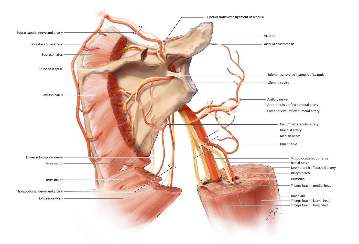
Arteries and Nerves of the Posterolateral Shoulder
Neurovascular structures in the posterior shoulder are complex and difficult to see during dissection. This graphic visualizes structures supplying the posterior muscles of the shoulder as well as their relationships to anterior structures.
Procreate; Adobe Illustrator
TOOLS

REFERENCES
Agur, A. M. R., & Dalley, A. F. (2017). Grant's atlas of anatomy (14th ed.). Philadelphia, PA: Wolters Kluwer.
Frank H. Netter (1989). Atlas of Human Anatomy. Summit, NJ: Ciba-Geigy.
KenHub. (2012). Biceps brachii muscle level. Retrieved April 19, 2021, from https://www.kenhub.com/en/study/cross-sections-biceps-brachii-muscle
National Library of Medicine. (1998). Visible Human Project CT Dataset, Male Shoulder. [CT Data DICOM file]. Retrieved from https://medicine.uiowa.edu/mri/facility-resources/images/visible-human-project-ct-datasets
Schuenke, Michael, Erik Schulte, and Udo Schumacher. (2014). Thieme Atlas of Anatomy (2nd ed.). New York, NY: Thieme Medical Publishers.